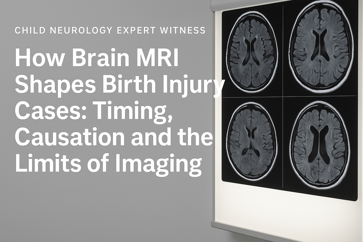
In birth injury and medical malpractice litigation, few tools are as critical as brain MRI scans when evaluating cases involving neonatal encephalopathy or hypoxic-ischemic encephalopathy (HIE). These conditions raise complex questions about timing of injury, causation, and long-term outcomes. Attorneys handling HIE cases must understand how MRI is used to assess newborn brain injury, what specific imaging patterns can reveal about the timing and severity of an event, and how expert witnesses interpret these findings in the context of standard of care. Properly understood, neonatal MRI can support or challenge key claims in litigation, making it a central component in both plaintiff and defense strategy.
What Can Neonatal MRI Reveal About the Timing of Brain Injury?
One of the most important aspects of MRI in HIE cases is its ability to help determine when the brain injury occurred. This is often a critical legal question: did the injury take place during labor and delivery, shortly after birth, or earlier in pregnancy?
Advanced MRI techniques such as diffusion-weighted imaging (DWI) and apparent diffusion coefficient (ADC) mapping can distinguish acute injuries from more chronic findings. For example, injury to the basal ganglia and thalamus is commonly associated with sudden, severe hypoxic events, while watershed injuries tend to reflect a longer period of partial oxygen deprivation. These findings can either support or challenge the argument that a preventable event occurred during delivery.
How Is MRI Used to Assess Severity and Predict Outcomes in HIE?
MRI is also essential for assessing the extent of brain injury, which has direct implications for expert analysis, damages and life care planning. Imaging is often performed in stages. The first scan is typically done between three and seven days after birth, when findings are most reliable. In some cases, additional imaging is conducted later to evaluate longer-term changes such as brain atrophy or delayed brain development.
MR spectroscopy (MRS), when available, adds another layer of insight by measuring brain metabolism. A high lactate-to-N-acetylaspartate (Lac/NAA) ratio in key brain regions is strongly associated with more severe injury and worse developmental outcomes.
What Are the Limitations of MRI in Birth Injury Cases?
Despite its importance, MRI has limitations that attorneys must be prepared to address. If performed too early, often within the first 24 hours of life, imaging may not show the full extent of the injury. Some types of brain damage, especially at the microscopic or metabolic level, may not be visible on conventional scans. Additionally, the accuracy of MRI findings depends on the quality of the scan, the imaging protocols used and the expertise of the interpreting radiologist.
In legal proceedings, these limitations can become focal points of disagreement between opposing experts. A normal early MRI does not necessarily rule out significant brain injury, and conversely, an abnormal scan may not always indicate that negligence occurred. This is why timing, clinical context and expert interpretation are so important.
How Do Experts Use MRI Findings in HIE Litigation?
Expert witnesses play a central role in interpreting MRI findings and explaining their implications to the court. Pediatric neurologists and neuroradiologists are commonly retained in these cases to analyze the scans, determine the likely timing and mechanism of injury, and assess how the imaging correlates with clinical symptoms and long-term prognosis.
These experts must be prepared to explain complex medical findings in clear, accessible language. Their testimony often hinges not only on what is visible in the imaging, but also on what the imaging does not show. For example, the absence of injury in a certain brain region may help rule out certain mechanisms, while the presence of specific patterns can support conclusions about the timing and severity of the hypoxic event.
How Can Attorneys Use MRI Strategically in Building or Defending a Case?
To make full use of MRI in litigation, attorneys should obtain complete imaging records, including raw DICOM files and technical details of each scan. Reviewing the imaging timeline in conjunction with the clinical record can uncover delays, omissions, or inconsistencies in care that may be relevant to the legal argument.
Early consultation with experienced medical experts is essential. These experts can help identify whether the imaging supports the alleged timing of injury, whether the scan protocols were appropriate, and whether the findings align with the child’s current neurological status. Attorneys can then use this analysis to inform their approach to depositions, settlement discussions, or trial presentation.
In court, annotated images and demonstrative exhibits can be powerful tools to help jurors understand the nature of the injury. However, these must be carefully developed and presented by experts who can stand up to scrutiny and explain their methodology clearly.
Why Does Understanding MRI Matter in HIE Litigation?
In cases involving neonatal encephalopathy and HIE, brain MRI is often one of the most objective and persuasive pieces of evidence available. It helps establish when and how the injury occurred, how severe it is, and what outcomes are likely. When interpreted by the right experts and presented effectively, MRI findings can strongly support claims related to causation and damages or it can expose weaknesses in those claims.
At the same time, attorneys must remain aware of MRI’s limitations and the potential for misinterpretation. Imaging should always be viewed in the context of the broader medical record, and conclusions should be grounded in accepted medical standards.
In summary, brain MRI serves as a foundational element in the evaluation and litigation of birth injury cases involving HIE. Attorneys who understand its value and constraints will be better prepared to advocate for their clients and navigate the medical complexity of HIE claims.
If you are working on a case involving neonatal encephalopathy or HIE and would benefit from a clinical perspective on the neuroimaging or neurologic findings, I welcome you to get in touch. I approach each review with the same care and objectivity I apply in clinical practice and teaching, with a focus on helping clarify complex medical questions in a legally relevant context.
